Abstract
Doppler echocardiography is commonly used for functional assessment of heart valves such as the mitral valve.
Currently, these measurements are performed manually — a laborious and subjective process.
This project demonstrates the feasibility of using convolutional neural networks (CNNs) to fully automate mitral inflow Doppler analysis.
Experiments show that the automated system yields performance comparable to clinical experts.
Introduction
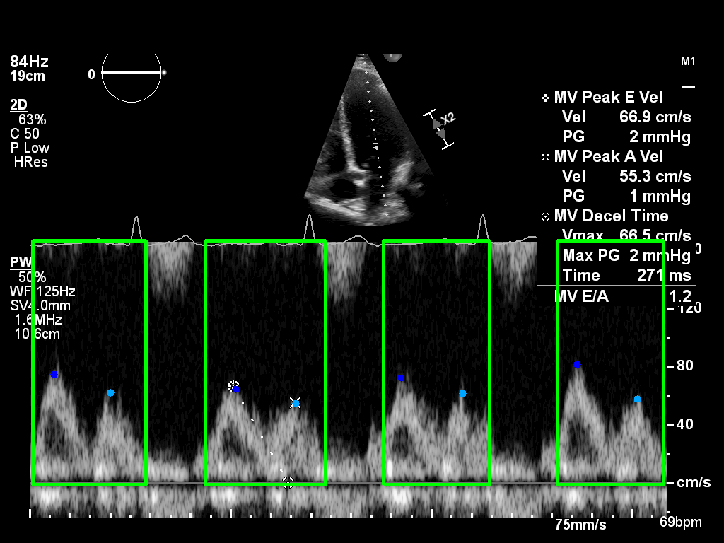
Transmitral Doppler echocardiography is a key technique for noninvasive assessment of diastolic function.
The Doppler velocity profile consists of two waves:
- E-wave — early ventricular filling
- A-wave — atrial contraction phase
Accurate measurement of these waves is critical for diagnosing diastolic dysfunction.
However, manual tracing introduces observer variability and consumes valuable clinical time.
Automating this process can help standardise measurement protocols, improve reproducibility, and enhance clinical workflow.
Method
A dataset of 500 echocardiographic examinations was obtained from Imperial College Healthcare NHS Trust’s echocardiogram database.
All studies were performed by experienced echocardiographers using GE and Philips ultrasound systems, under ethical approval (IRAS ID: 243023).
Expert cardiologists manually annotated E- and A-wave peaks, and DICOM-formatted Doppler images were zero-padded to 1024×1024 px.
Data was split into 400 training and 100 testing cases.
The proposed deep learning pipeline includes two stages:
- Heartbeat Detection: Using a Faster-RCNN object detection model to localise individual beats.
- Landmark Detection: Employing a Convolutional Pose Machine-based network (Wei et al., 2016) to identify E- and A-wave peaks.
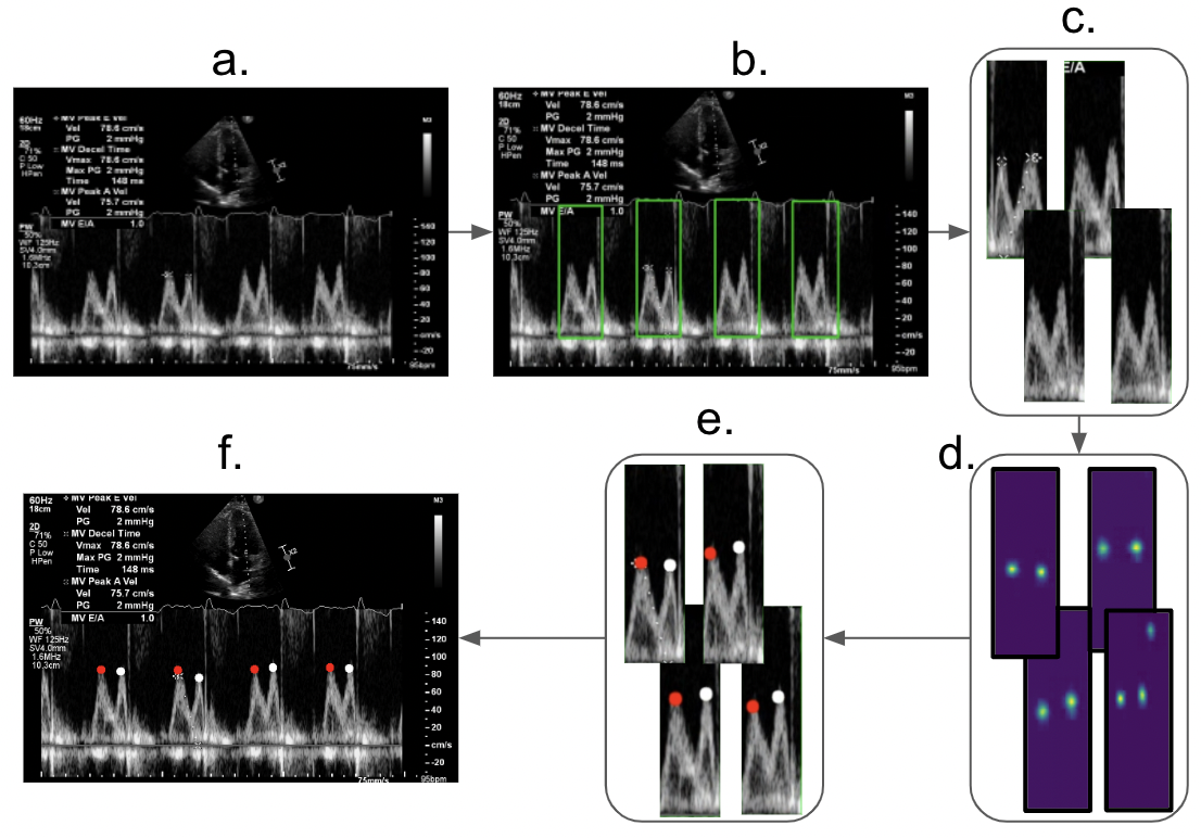 Figure 1: End-to-end pipeline for automatic Doppler inflow analysis.
The models were implemented in TensorFlow and trained on an NVIDIA RTX 3090 GPU.
Post-processing included non-maximum suppression for bounding boxes and heatmap decoding for wave peak extraction.
Results
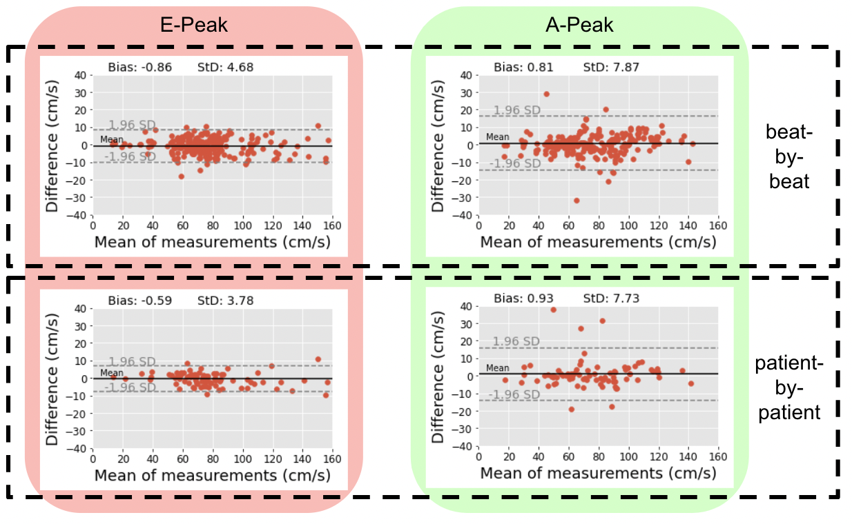 Figure 2: Bland–Altman plots comparing automated and manual measurements.
Bias between automated and manual measurements was < 1 cm/s for both E- and A-waves.
The A-wave showed slightly greater variability, reflecting its more complex morphology.
These results confirm that the CNN-based system performs on par with human experts, supporting potential integration into echocardiographic analysis workflows.
All datasets, trained models, and code developed in this study are freely available for benchmarking and research use.
Dataset Access
To request access to the Mitral Inflow Doppler (MInD) dataset, please complete the dataset request form below:
Project Team
Reference
Wei, S.-E., Ramakrishna, V., Kanade, T., & Sheikh, Y. (2016).
Convolutional Pose Machines. CVPR 2016, pp. 4724–4732.