Echo View Classification
Start Labelling →
Project Overview
We are training an AI model for detailed classification of echocardiographic views , covering 47 distinct categories .apical 3-chamber and apical 4-chamber — can appear similar, making it difficult to determine the correct label.
Please examine each image carefully to distinguish between these views.not fit any available category , select “None of the above” .“Not sure” .
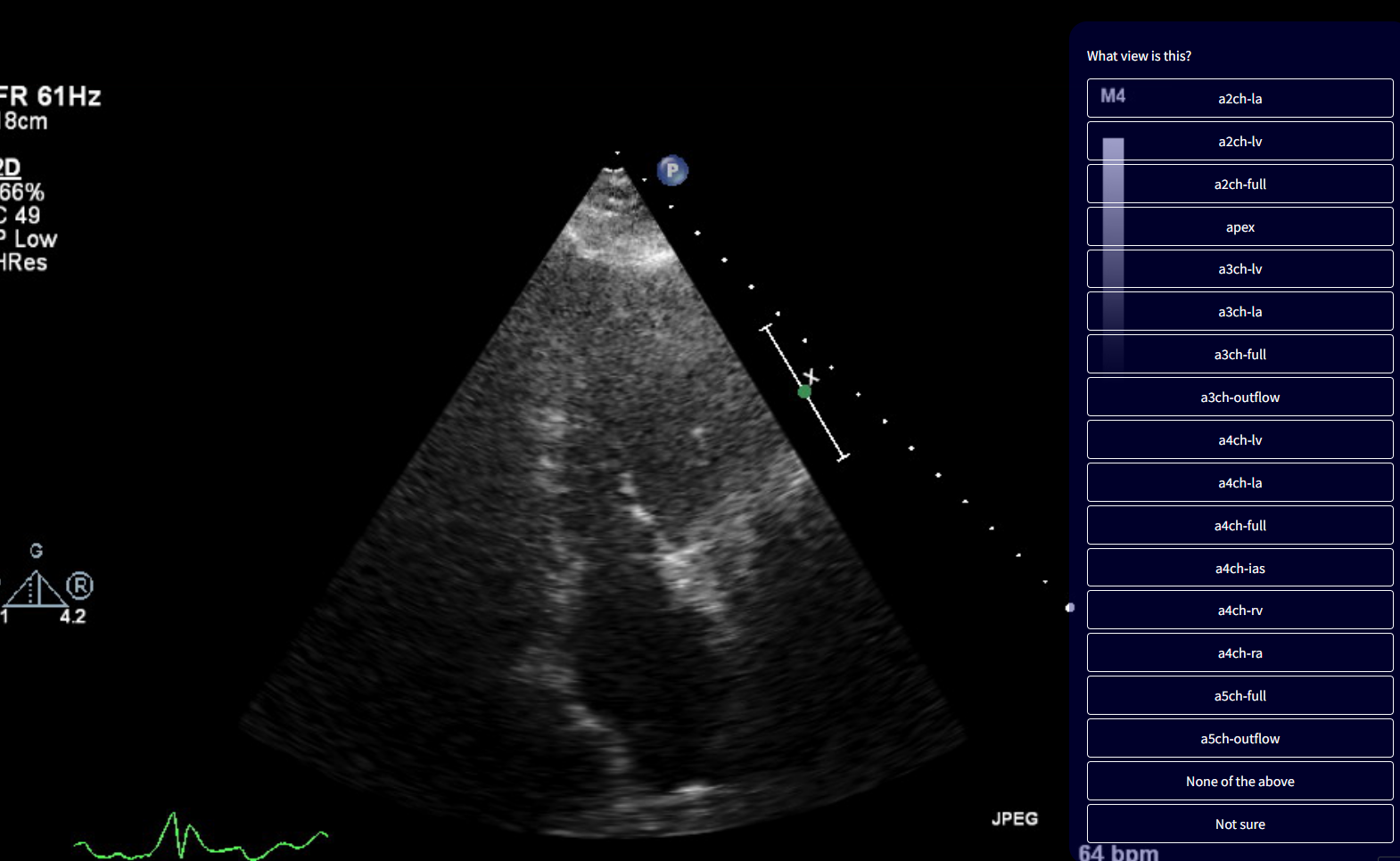
Visual Example
Instructional Video
Watch a 1-minute demo video
VIDEO
Start Labelling
View Descriptions
Apical Views
View
Description
a2ch-la
Apical 2-chamber, focused on the left atrium
a2ch-lv
Apical 2-chamber, focused on the left ventricle
a2ch-full
Apical 2-chamber
apex
Any apical window, focused on the apex
a3ch-la
Apical 3-chamber, focused on the left atrium
a3ch-lv
Apical 3-chamber, focused on the left ventricle
a3ch-full
Apical 3-chamber
a3ch-outflow
Apical 3-chamber, focused on the aortic valve
a4ch-la
Apical 4-chamber, focused on the left atrium
a4ch-lv
Apical 4-chamber, focused on the left ventricle
a4ch-full
Apical 4-chamber
a4ch-ias
Apical 4-chamber, focused on the inter-atrial septum
a4ch-ra
Apical 4-chamber, focused on the right atrium
a4ch-rv
Apical 4-chamber, focused on the right ventricle
a5ch-full
Apical 5-chamber
a5ch-outflow
Apical 5-chamber, focused on the aortic valve
Doppler Views
View
Description
doppler-ao-descending
Spectral Doppler of the descending aorta
doppler-mv
Spectral Doppler of the mitral valve
doppler-av
Spectral Doppler of the aortic valve
doppler-pv
Spectral Doppler of the pulmonary valve
doppler-tv
Spectral Doppler of the tricuspid valve
doppler-tissue-lateral
Tissue Doppler of the left ventricular lateral wall
doppler-tissue-septal
Tissue Doppler of the left ventricular septal wall
doppler-tissue-rv
Tissue Doppler of the right ventricular free wall
PLAX Views
View
Description
plax-full-out
Parasternal long-axis, zoomed out
plax-full-lv
Parasternal long-axis, focused on the left ventricle
plax-full-la
Parasternal long-axis, focused on the left atrium
plax-full-rv-ao
Parasternal long-axis, focused on the right ventricle and aorta
plax-full-mv
Parasternal long-axis, centered on the mitral valve
plax-valves-av
Parasternal long-axis, focused on the aortic valve
plax-valves-mv
Parasternal long-axis, focused on the mitral valve
plax-tv
Parasternal inflow view including tricuspid valve
PSAX Views
View
Description
psax-all
Parasternal short-axis, valve level, including all valves
psax-av
Parasternal short-axis, focused on aortic valve
psax-tv
Parasternal short-axis, focused on tricuspid valve
psax-pv
Parasternal short-axis, focused on pulmonary valve
psax-lv-base
Parasternal short-axis, left ventricle base level
psax-lv-mid
Parasternal short-axis, left ventricle mid-level
psax-lv-apex
Parasternal short-axis, left ventricle apex level
M-mode Views
View
Description
mmode-a4ch-rv
M-mode for measuring TAPSE
mmode-ivc
M-mode of the inferior vena cava
mmode-plax-mitral
M-mode of the mitral valve in the parasternal long-axis
mmode-plax-av
M-mode of the aortic valve in the parasternal long-axis
mmode-plax-lv
M-mode of the left ventricle in the parasternal long-axis
Subcostal / Suprasternal Views
View
Description
subcostal-heart
Subcostal window, focused on the heart
subcostal-ivc
Subcostal window, focused on the inferior vena cava
suprasternal
Suprasternal view