UWL Centre for Translational Healthcare Research
Download Dataset Trained Model References
TTE47 is a dataset derived from a random sample of real-world echocardiographic studies collected at Imperial College Healthcare NHS Trust, comprising a total of 91,139 images. Ethical approval for the study was granted by the Health Research Authority (Integrated Research Application System IRAS identifier: 243023). Only studies with complete demographic information and without intravenous contrast were included.
Each image was manually annotated by a cardiologist (“Expert 1”) via a web-based platform (https://unityimaging.net/) and assigned to one of 47 predefined categories. The dataset was divided into training (76,589), validation (9,103), and test (5,447) sets, with no patient or study overlap between splits. The echocardiograms were obtained from 19,169 unique studies.
Two additional clinical experts independently annotated the test set. These experts were blinded to the original labels and to each other’s annotations. Unlike Expert 1, who was required to assign exactly one label to each image, Experts 2 and 3 were allowed to select “not sure” when appropriate. The test set was intentionally sized to ensure reliable evaluation while remaining feasible for detailed multi-expert annotation across all 47 views.
The test subset, containing 5,447 images with annotations from all three experts, is released under the Creative Commons Attribution-NonCommercial-ShareAlike 4.0 International license. Release of the dataset was approved by the South Central – Oxford C Research Ethics Committee (IRAS ID: 279328, REC reference: 20/SC/0386). Due to data governance restrictions, the training set cannot be made public, but pre-trained and fine-tuned model weights are provided.
Demographic profile of the TTE47 dataset, encompassing a diverse adult population with representation across multiple age groups, BMI categories, and sexes.
| Characteristic | Category | Proportion (%) |
|---|---|---|
| Age (years) | 18–30 | 2.3 |
| 31–50 | 12.7 | |
| 51–70 | 27.9 | |
| 71+ | 57.2 | |
| Sex | Female | 50.5 |
| Male | 48.6 | |
| Other | 0.8 | |
| BMI (kg/m²) | 18–24.9 (Normal) | 36.0 |
| 25–29.9 (Overweight) | 34.9 | |
| 30–34.9 (Obesity I) | 18.0 | |
| 35+ (Obesity II+) | 11.1 |
Distribution of echocardiographic view classes across the training, validation, and test sets in TTE47.
| View | Training | Validation | Testing | Total |
|---|---|---|---|---|
| a2ch-full | 2931 | 349 | 209 | 3489 |
| a2ch-la | 1936 | 230 | 138 | 2304 |
| a2ch-lv | 2987 | 355 | 213 | 3555 |
| a3ch-full | 3561 | 424 | 254 | 4239 |
| a3ch-la | 1622 | 193 | 115 | 1930 |
| a3ch-lv | 1926 | 229 | 137 | 2292 |
| a3ch-outflow | 637 | 75 | 45 | 757 |
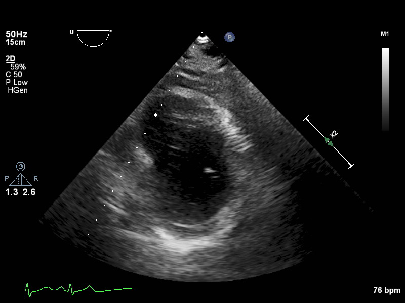
| a4ch-full | 3353 | 399 | 239 | 3991 |
| a4ch-ias | 1319 | 157 | 94 | 1570 |
| a4ch-la | 2733 | 325 | 195 | 3253 |
| a4ch-lv | 2495 | 297 | 178 | 2970 |
| a4ch-ra | 1713 | 204 | 122 | 2039 |
| a4ch-rv | 1236 | 147 | 88 | 1471 |
| a5ch-full | 753 | 89 | 53 | 895 |
| a5ch-outflow | 786 | 93 | 56 | 935 |
| apex | 723 | 86 | 51 | 860 |
| doppler-ao | 1489 | 177 | 106 | 1772 |
| doppler-av | 2587 | 308 | 184 | 3079 |
| doppler-mv | 1701 | 202 | 121 | 2024 |
| doppler-pv | 1776 | 211 | 126 | 2113 |
| doppler-tissue-lateral | 1120 | 133 | 80 | 1333 |
| doppler-tissue-rv | 780 | 92 | 55 | 927 |
| doppler-tissue-septal | 1034 | 123 | 73 | 1230 |
| doppler-tv | 1902 | 226 | 135 | 2263 |
| mmode-a4ch-rv | 1592 | 189 | 113 | 1894 |
| mmode-ivc | 1159 | 138 | 82 | 1379 |
| mmode-plax-av | 1374 | 163 | 98 | 1635 |
| mmode-plax-lv | 492 | 58 | 35 | 585 |
| mmode-plax-mitral | 768 | 91 | 54 | 913 |
| plax-full-la | 1123 | 133 | 80 | 1336 |
| plax-full-lv | 1799 | 214 | 128 | 2141 |
| plax-full-mv | 1307 | 155 | 93 | 1555 |
| plax-full-out | 1509 | 179 | 107 | 1795 |
| plax-full-rv-ao | 961 | 114 | 68 | 1143 |
| plax-tv | 1688 | 201 | 120 | 2009 |
| plax-valves-av | 2000 | 238 | 142 | 2380 |
| plax-valves-mv | 1841 | 219 | 131 | 2191 |
| psax-all | 2097 | 249 | 149 | 2495 |
| psax-tv | 1335 | 159 | 95 | 1589 |
| psax-av | 2125 | 253 | 151 | 2529 |
| psax-pv | 1868 | 222 | 133 | 2223 |
| psax-lv-base | 1437 | 171 | 102 | 1710 |
| psax-lv-mid | 1669 | 198 | 119 | 1986 |
| psax-lv-apex | 1627 | 193 | 116 | 1936 |
| subcostal-heart | 1212 | 144 | 86 | 1442 |
| subcostal-ivc | 849 | 101 | 60 | 1010 |
| suprasternal | 1657 | 197 | 118 | 1972 |
| Total | 76589 | 9103 | 5447 | 91139 |
Below are 6 major categories. Click a category to reveal its table of TTE47 views.
| Image | View Name | Description |
|---|---|---|
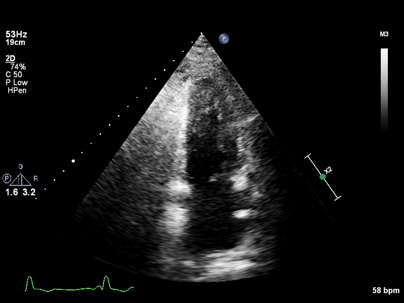 |
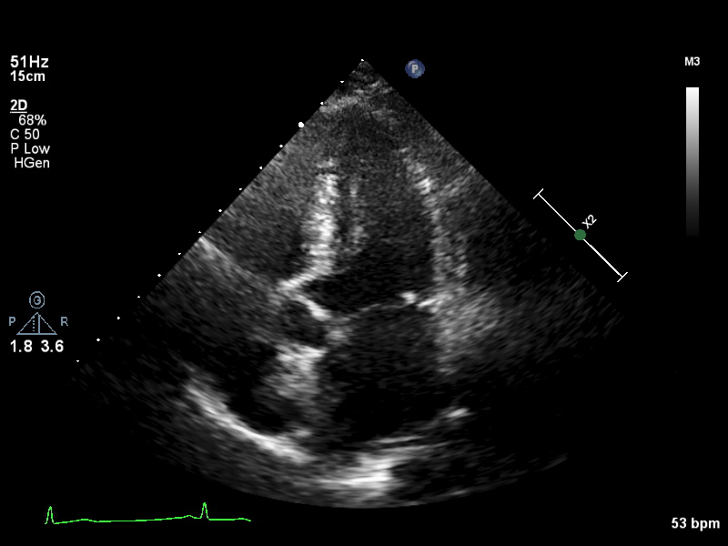
a2ch-full |
Apical 2-chamber, covering the depth of the whole of the LV and LA |
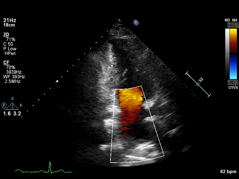 |
a2ch-la |
A2CH with Doppler near MV/LA or 2D not covering full LV depth |
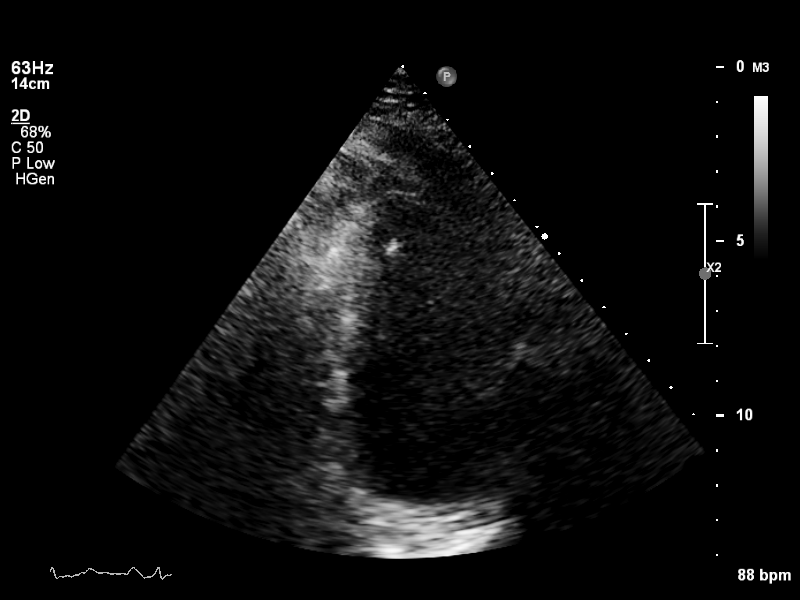 |
a2ch-lv |
A2CH covering the whole depth of the LV, but not the whole depth of the LA |
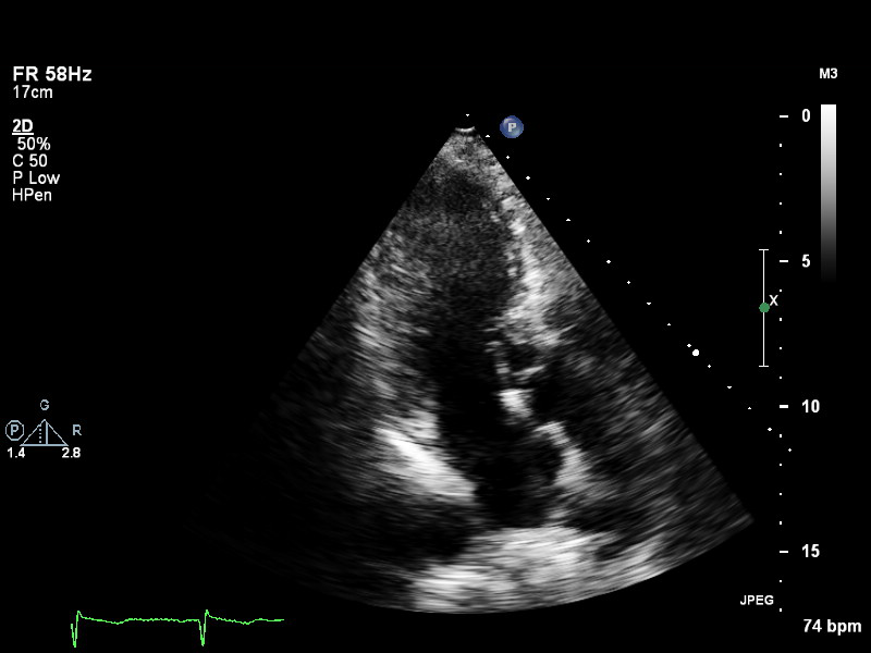 |
a3ch-full |
Apical 3-chamber, covering the whole depth of the LV and the LA |
 |
a3ch-la |
A3CH with Doppler near MV/LA or 2D not covering full LV |
 |
a3ch-lv |
A3CH covering the whole depth of the LV, but not the whole depth of the LA |
 |
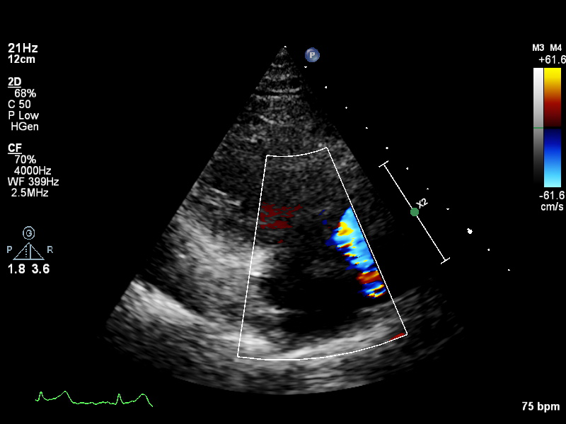
a3ch-outflow |
A3CH emphasizing LVOT (colour focused on LVOT or limited b&w of LVOT area) |
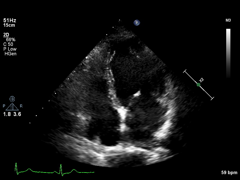 |
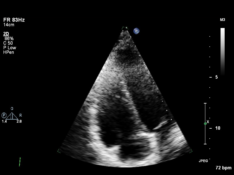
a4ch-full |
Apical 4-chamber showing the full depth of all four chambers (no LV/RV focus) |
 |
a4ch-ias |
Apical 4-chamber, focused on the inter-atrial septum |
 |
a4ch-la |
A4CH with Doppler near MV/LA or 2D covering full LA, not full LV |
 |
a4ch-lv |
A4CH covering the whole depth of the LV, not the whole depth of the LA |
 |
a4ch-ra |
Apical 4-chamber focused on the right atrium, or colour Doppler on TV/RA |
 |
a4ch-rv |
Apical 4-chamber focused on the right ventricle |
 |
a5ch-full |
Apical 5-chamber from apex to back of the atria, no LVOT colour |
 |
a5ch-outflow |
A5CH including LVOT; may lack full depth or has LVOT colour |
 |
apex |
Any apical window whose depth is insufficient to reach the mitral ring |
| Image | View Name | Description |
|---|---|---|
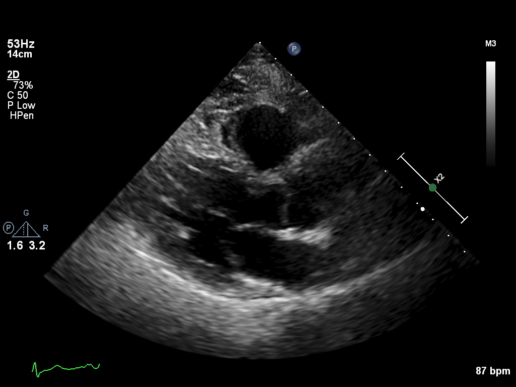
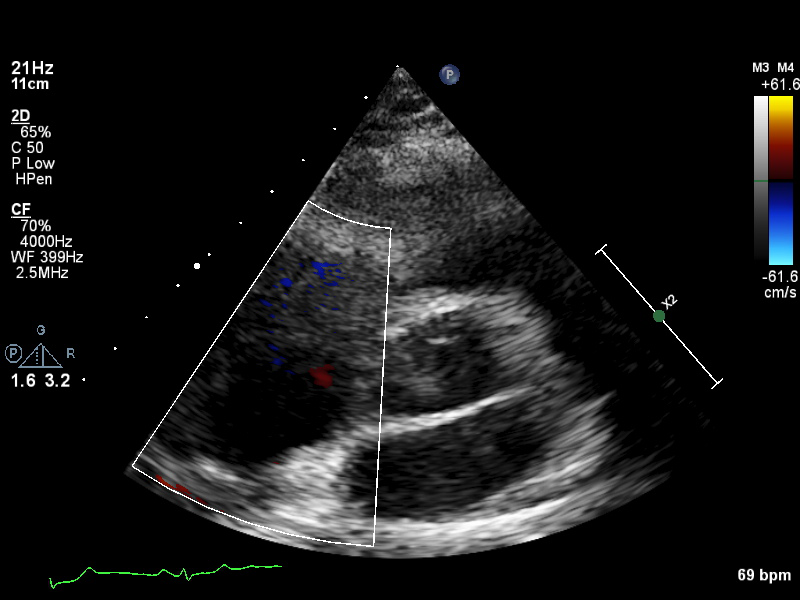
 |
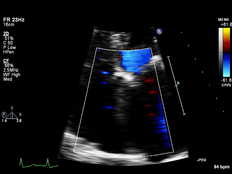
plax-full-la |
Parasternal long-axis, sector covers whole LA; intended for LA measurements |
 |
plax-full-lv |
PLAX with imaging sector covering LV but not the whole LA |
 |
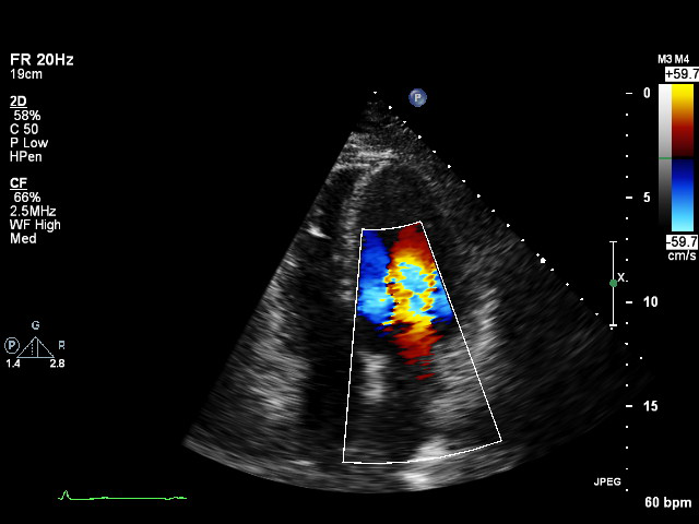
plax-full-mv |
PLAX centered on MV (heavy zoom losing LA/LV parts or MV colour) |
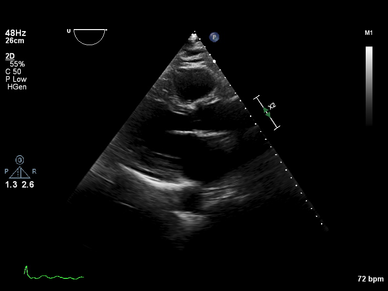 |
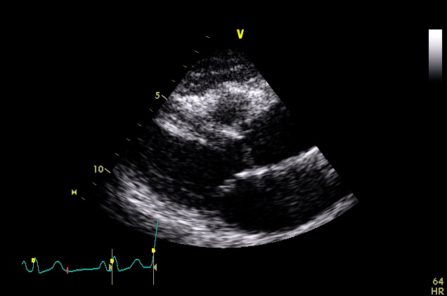
plax-full-out |
Zoomed-out PLAX |
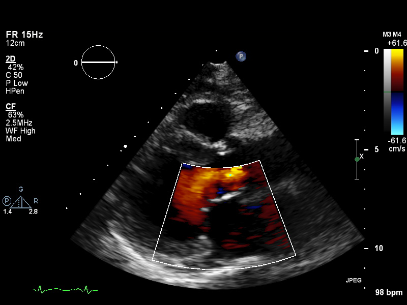
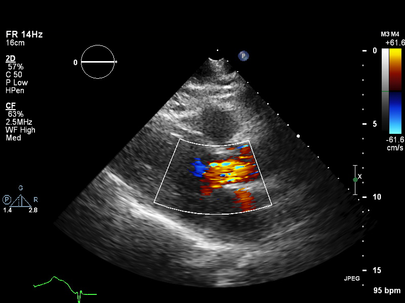 |
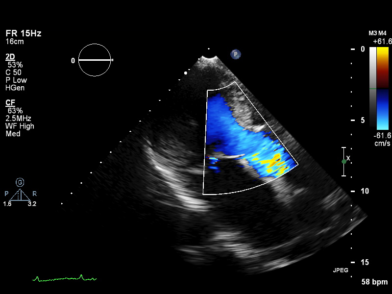
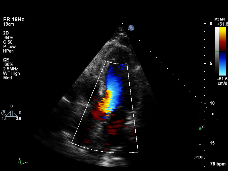
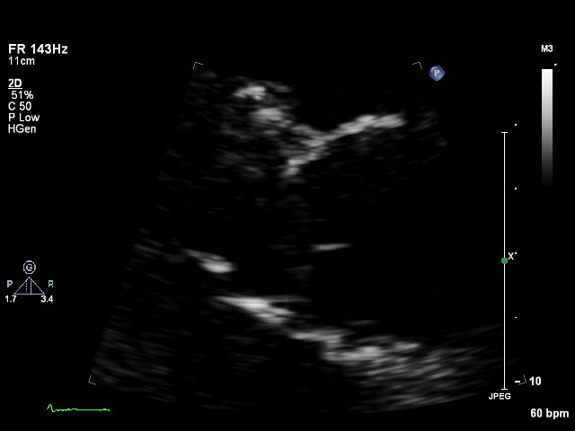
plax-full-rv-ao |
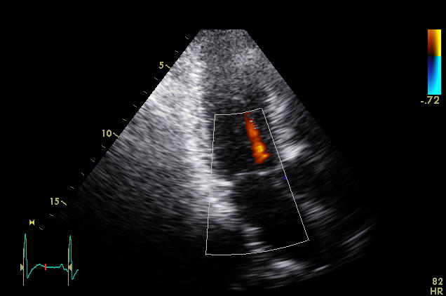
Focus on RV & aorta or colour Doppler over RV/AV/Aorta |
 |
plax-tv |
Parasternal inflow view including tricuspid valve |
 |
plax-valves-av |
PLAX focused on the aortic valve |
 |
plax-valves-mv |
PLAX focused on the mitral valve |
| Image | View Name | Description |
|---|---|---|
 |
psax-all |
Valve level, field of view covers TV/AV/PV positions |
 |
psax-tv |
Focused on tricuspid valve |
 |
psax-av |
Focused on aortic valve |
 |
psax-pv |
Focused on pulmonary valve |
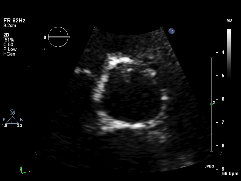
 |
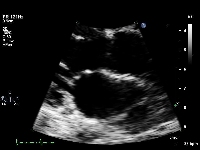
psax-lv-base |
Left ventricle base level |
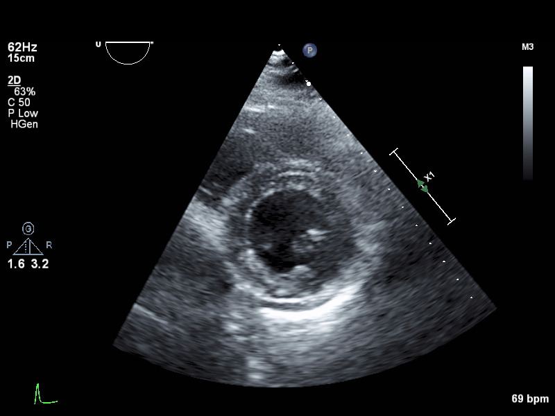 |
psax-lv-mid |
Left ventricle mid level |
 |
psax-lv-apex |
Left ventricle apex level |
| Image | View Name | Description |
|---|---|---|
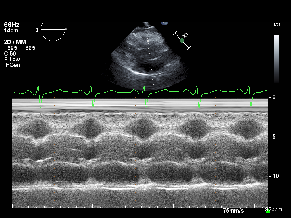
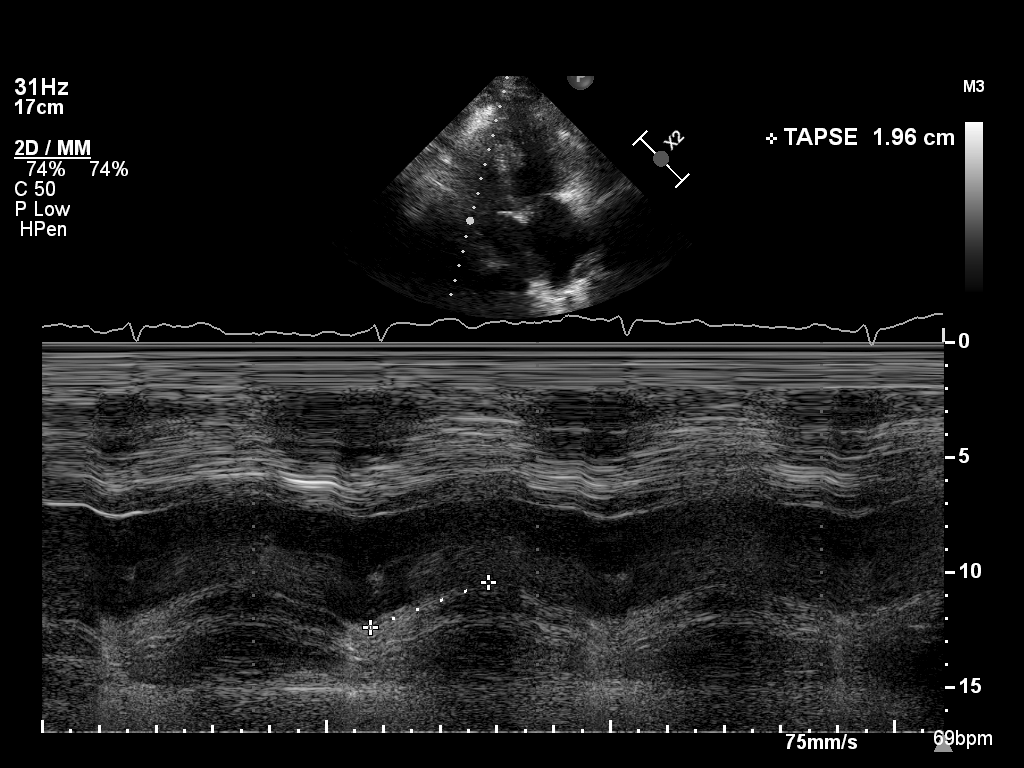 |
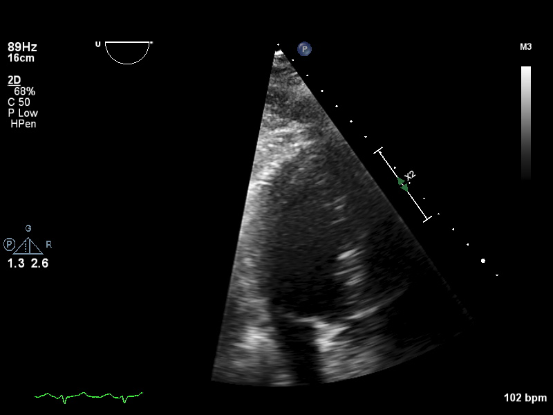
mmode-a4ch-rv |
M-mode for measuring TAPSE |
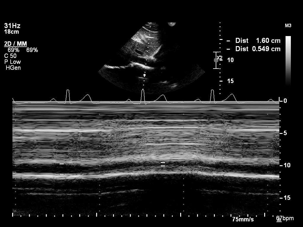 |
mmode-ivc |
M-mode of the inferior vena cava |
 |
mmode-plax-av |
M-mode of the aortic valve in PLAX |
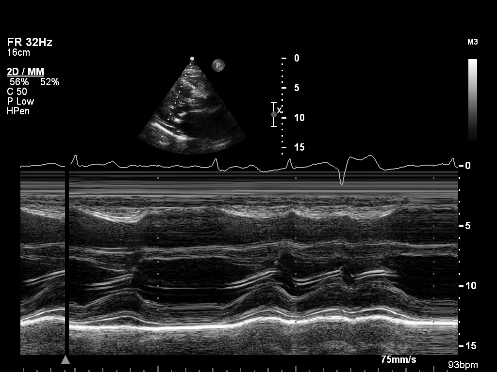 |
mmode-plax-lv |
M-mode in PLAX focused on LV walls |
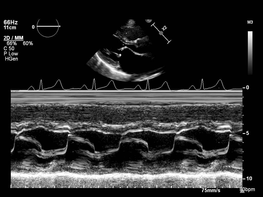 |
mmode-plax-mitral |
M-mode in PLAX focused on the mitral valve |
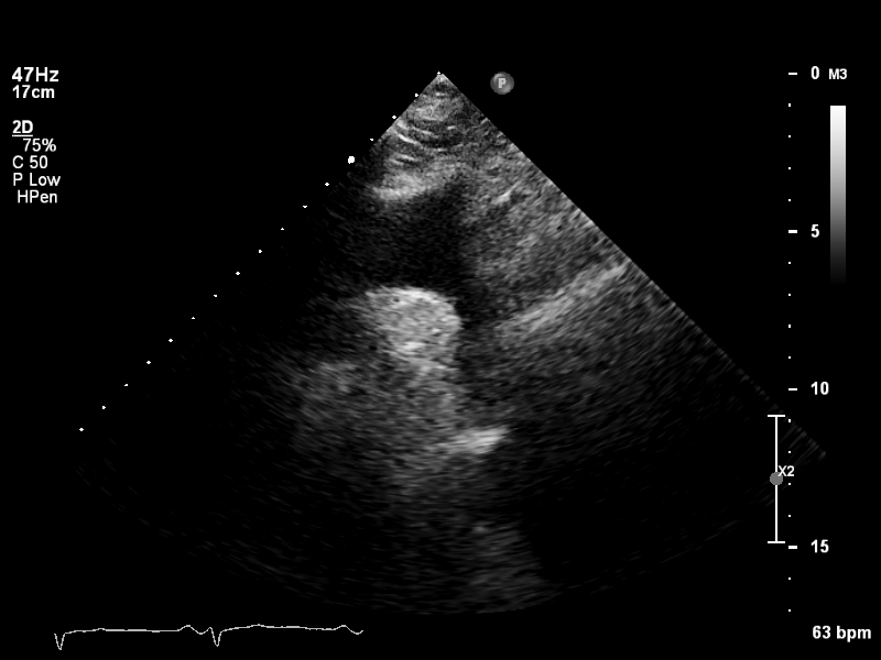
| Image | View Name | Description |
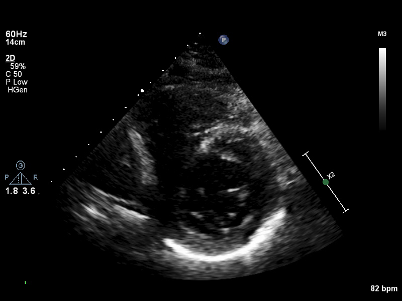
|---|---|---|
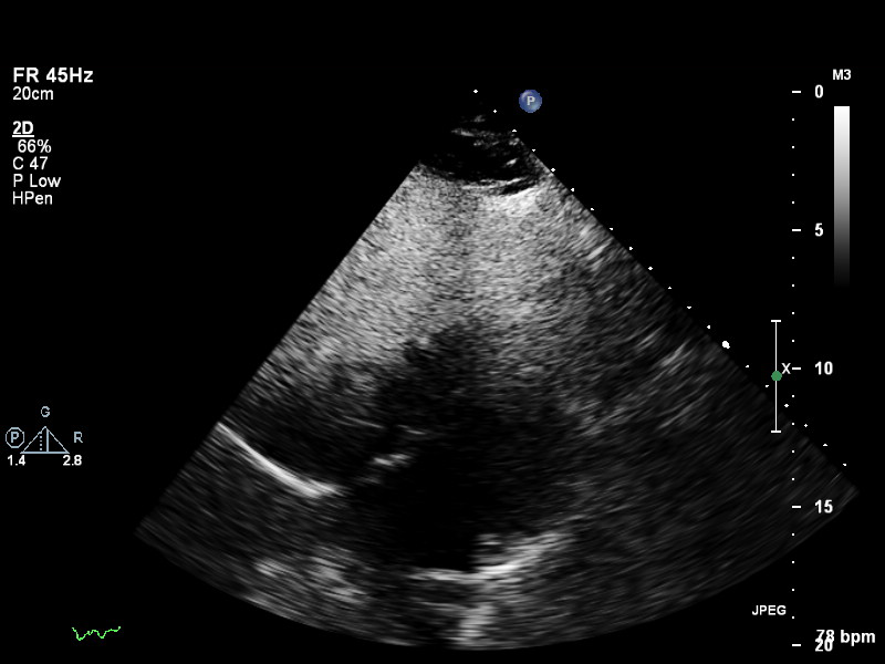 |
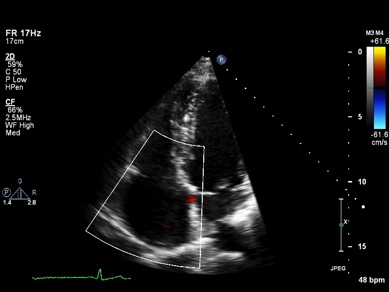
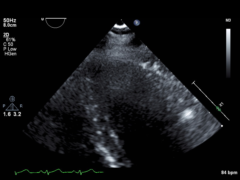
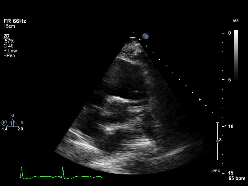
subcostal-heart |
Subcostal window, focused on the heart |
 |
subcostal-ivc |
Subcostal window, focused on the inferior vena cava |
 |
suprasternal |
Suprasternal view |
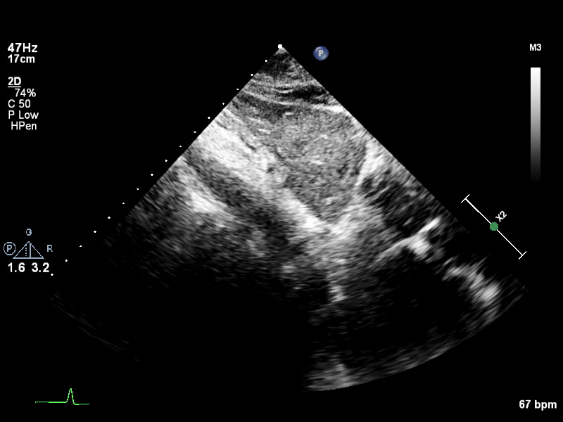
| Image | View Name | Description |
|---|---|---|
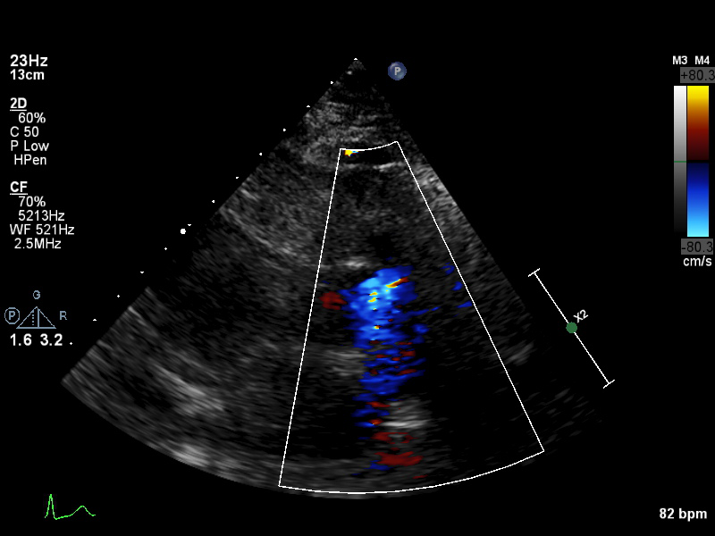
 |
doppler-ao |
Spectral Doppler of the descending aorta |
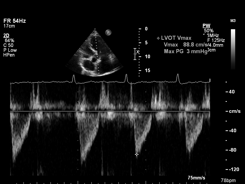 |
doppler-av |
Spectral Doppler of the aortic valve |
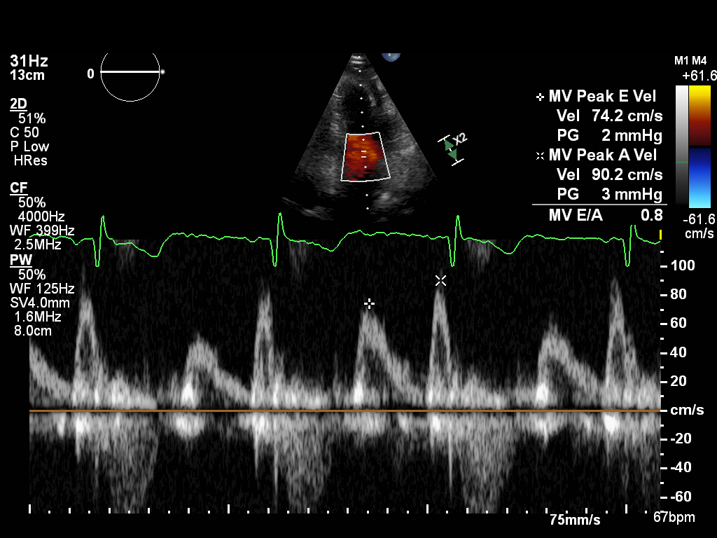 |
doppler-mv |
Spectral Doppler of the mitral valve |
 |
doppler-pv |
Spectral Doppler of the pulmonary valve |
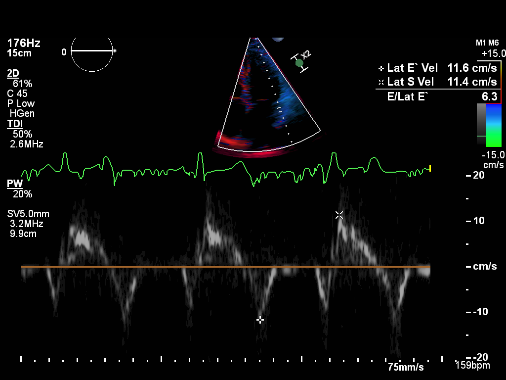 |
doppler-tissue-lateral |
Tissue Doppler of the LV lateral wall |
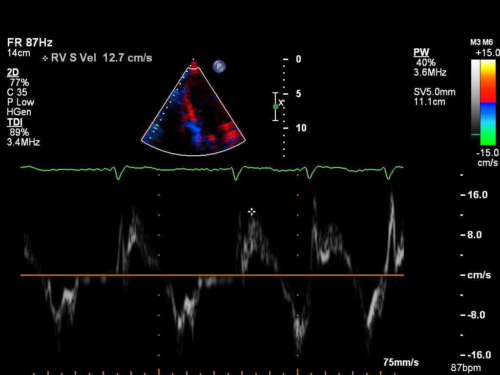 |
doppler-tissue-rv |
Tissue Doppler of the RV free wall |
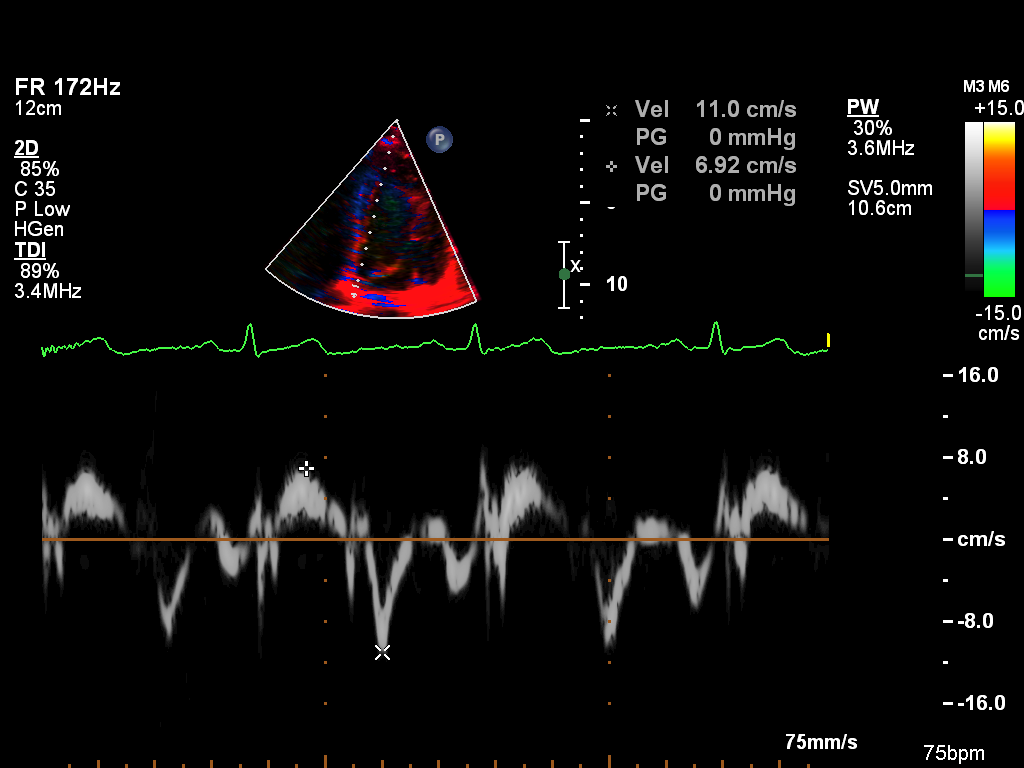 |
doppler-tissue-septal |
Tissue Doppler of the LV septal wall |
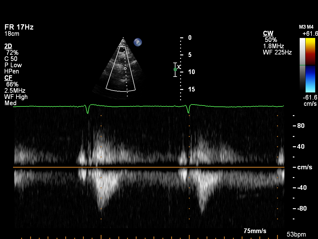
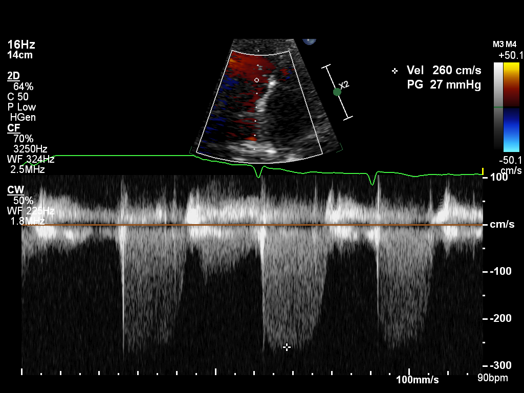 |
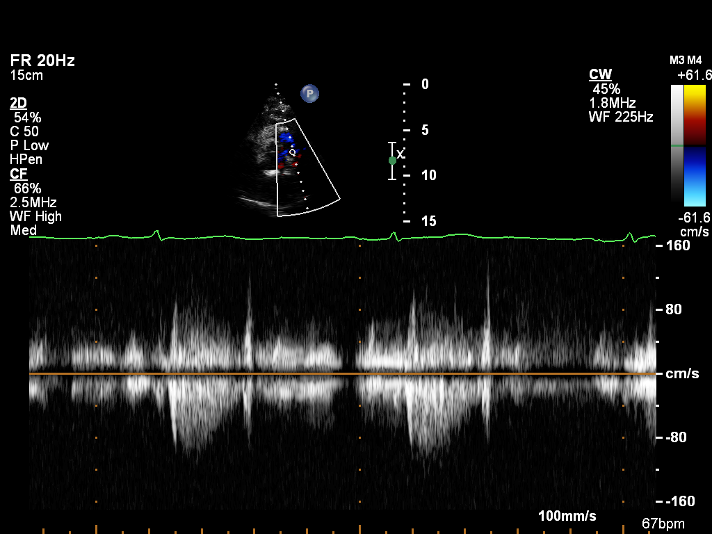
doppler-tv |
Spectral Doppler of the tricuspid valve |
If you wish to request access, please complete the form below: