UWL Centre for Translational Healthcare Research
The UNITY-ROT dataset is a publicly available benchmark for assessing rotational misalignment in apical four-chamber (A4C) echocardiographic images. The dataset was curated from real-world transthoracic echocardiography studies acquired at Imperial College Healthcare NHS Trust under Health Research Authority approval for the anonymised export of clinical imaging data.
The source database contains heterogeneous echocardiographic studies acquired across a broad patient population using ultrasound systems from multiple vendors. This diversity was intentionally preserved, as rotational misalignment is influenced by patient anatomy, acoustic windows, probe positioning, acquisition protocols, and operator variability. The aim of this dataset is therefore to reflect the variability encountered in routine clinical echocardiography rather than a narrowly curated set of idealised examples.
Studies were acquired by experienced sonographers following standard clinical imaging protocols. All patient-identifiable metadata were removed prior to export. Ethical approval was obtained for the anonymised use of the imaging data originally collected during routine clinical care.
The UNITY-ROT benchmark was designed to support reproducible evaluation of AI systems for echocardiographic rotational alignment assessment. The dataset includes expert-ranked images and independent multi-expert evaluation sets to facilitate robust benchmarking of clockwise–anti-clockwise alignment prediction models.
The dataset and associated annotations are intended to support future research into echocardiographic quality assessment, representation learning, ranking-based supervision, and clinically meaningful evaluation of alignment models.
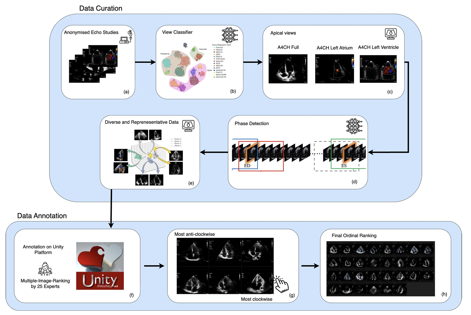
Candidate apical-view videos were first identified using an automated echocardiographic view classification framework. Three A4C-related subview categories were retained for this study:
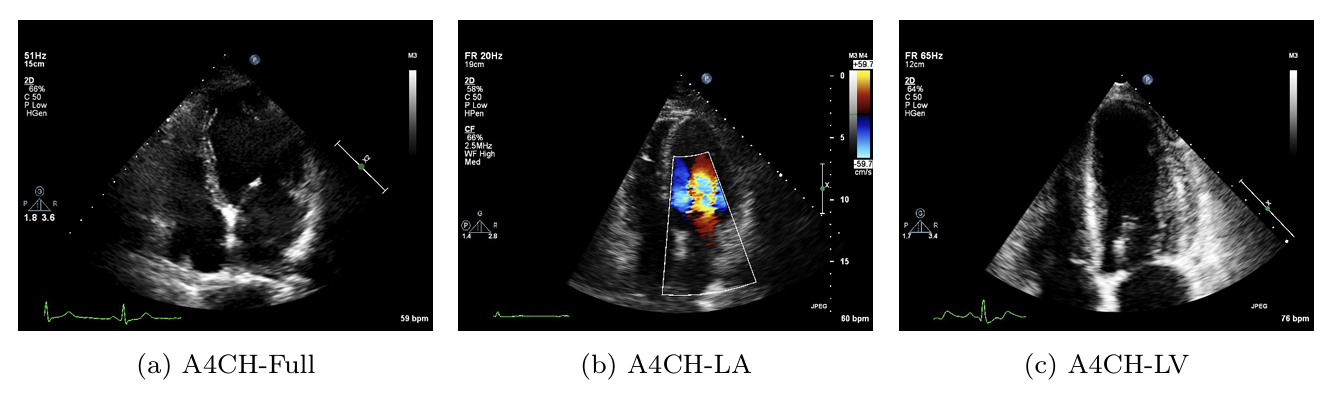
Representative examples of the three retained A4C-related subview categories are shown above.
A4CH-Full contains the complete four-chamber view, while A4CH-LA and A4CH-LV focus predominantly on the left atrium and left ventricle respectively.
Each video contained between one and three cardiac cycles. Representative frames were automatically selected using a cardiac phase detection model. For every video:
This resulted in four representative frames per video, capturing cardiac morphology across different phases of the cardiac cycle.
To prioritise informative examples and reduce redundant annotation effort, a diversity-based sample selection strategy was applied. Features extracted from the representative frames were aggregated into video-level embeddings using a CNN feature extractor. Principal Component Analysis (PCA) followed by clustering was then used to identify representative videos spanning the observed appearance distribution.
The most visually diverse and representative cases were prioritised for expert annotation.
The annotation protocol was designed to capture rotational alignment as a relative ranking problem rather than as an isolated absolute score.
Following diversity-based selection:
This produced a total of 400 videos for annotation.
Four representative frames were extracted from each video, yielding 1,600 images for expert annotation.
The images were annotated by a pool of 25 clinical experts using our custom-built web-based annotation platform:
Experts were presented with randomly shuffled batches of six images and asked to reorder them from:
according to perceived A4C rotational alignment.
This drag-and-drop multiple-image ranking strategy enabled experts to directly compare images instead of assigning isolated numerical scores. Relative ranking was selected because it provides robust consensus generation while reducing inter-observer variability.
The Unity platform dynamically updated four internal variables throughout the annotation process:
Images with low annotation counts or high disagreement were prioritised for further review until sufficient consensus was achieved.
Expert annotations were aggregated to produce:
The annotated development dataset comprised:
The dataset was divided into three non-overlapping subsets:
To prevent data leakage, images originating from the same echocardiographic study were restricted to a single subset.
For final evaluation against expert consensus, three additional independent evaluation sets were curated.
Each evaluation set contained:
Each set was independently ranked by 10 clinical experts, who reordered the images from most anti-clockwise to most clockwise according to perceived rotational alignment.
These independent expert rankings were aggregated into consensus rankings for benchmarking model performance against human expert agreement.
This evaluation strategy provides a clinically meaningful assessment of rotational alignment prediction models.
Clinical experts annotated images using the Unity Imaging platform through a drag-and-drop ranking interface. Images were presented in shuffled batches and reordered from most anti-clockwise to most clockwise according to perceived rotational alignment.
The annotation workflow and ranking strategy are illustrated in Figure 2.
The dataset package available for download includes:
Benchmark evaluation results and pretrained models will be released alongside the public dataset release.
If you wish to request access, please complete the form below: